General overview
Acanthamoeba spp. are free-living amoeba (FLA) that have been commonly found in freshwater, tap water, and recreational water. Acanthamoeba spp. are capable of causing a variety of infections, including Acanthamoeba keratitis, an eye infection that has been associated with contect lens usage and cornea damage, and Granulomatous Amebic Encephalitis (GAE), a serious central nervous system infection that primarily affects immunocompromised individuals (Marciano-Cabral & Cabral, 2003; Visvesvara, Moura, & Schuster, 2007)[2] . Considering these infections, some exposure routes of concern for Acanthamoeba spp. are the corneal and intranasal exposure routes.
Dean et al. (2020) fit dose response models to data from previously conducted animal studies for Acanthamoeba spp. and the corneal and instranasal exposure routes. These models are an important step towards characterizing the risk associated with FLA like Acanthamoeba for drinking water-relevant exposure scenarios. More detailed descriptions of the datasets, fitting methods, model evaluation, and results can be found in the published article: http://dx.doi.org/10.1111/risa.13603
Recommended Model
For the corneal exposure route, two dose response models are provided for consideration.
Marciano-Cabral, F., & Cabral, G. (2003). Acanthamoeba spp. as agents of disease in humans. Clinical Microbiology Reviews. https://doi.org/10.1128/CMR.16.2.273-307.2003
| ID | Exposure Route | # of Doses | Agent Strain | Dose Units | Host type | Μodel | LD50/ID50 | Optimized parameters | Response type | Reference |
|---|---|---|---|---|---|---|---|---|---|---|
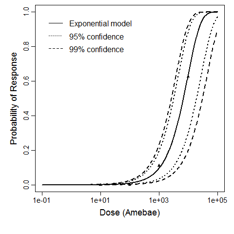
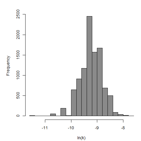
| Acanth_Cornea1 | eyes-cornea | 4.00 | Acanthamoeba Ac118 | no of trophozoites | rat | exponential | 6,886 | k = 1.01E-04 | infection |
Badenoch, P. R., Johnson, A. M., Christy, P. E., & Coster, D. J. (1990). Pathogenicity of Acanthamoeba and Corynebacterium in the Rat Cornea. Archives of Ophthalmology, 108, 1. Retrieved from https://jamanetwork.com/journals/jamaophthalmology/article-abstract/638228 |
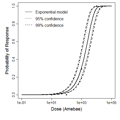
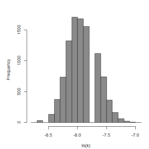
| Acanth_Cornea2 | eyes-cornea | 4.00 | Acanthamoeba Ac118 | no of trophozoites | rat | exponential | 1.91E+03 | k = 3.63E-04 | infection |
Badenoch, P. R., Johnson, A. M., Christy, P. E., & Coster, D. J. (1990). Pathogenicity of Acanthamoeba and Corynebacterium in the Rat Cornea. Archives of Ophthalmology, 108, 1. Retrieved from https://jamanetwork.com/journals/jamaophthalmology/article-abstract/638228 |
Badenoch et al. (1990) studied the combined effect of Acanthamoeba Ac118 (a group III isolate) and the bacterium Corynebacterium xerosis on the corneas of female Porton rats. A constant dose of 104 C. xerosis with increasing doses of Acanthamoeba spp. were injected into incisions in the rat corneas using a microsyringe (Badenoch et al. 1990).
The exponential model provided the best fit to the data.
Badenoch et al. (1990) studied the combined effect of Acanthamoeba Ac118 (a group III isolate) and the bacterium Corynebacterium xerosis on the corneas of female Portion rats. A constant dose of 106 C. xerosis with increasing doses of Acanthamoeba spp. were injected into incisions in the rat corneas using a microsyringe.
The exponential model provided the best fit to the data.


